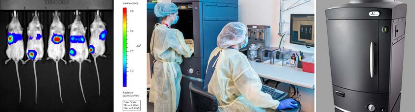
Bioluminescence and fluorescence optical imaging (BLI and FLI) is performed on two PerkinElmer IVIS® Spectrum Preclinical In Vivo Imaging Systems equipped with dedicated isoflurane anesthesia systems and a temperature controlled chamber. The IVIS® Spectrum in vivo imaging system uses leading optical imaging technology to facilitate non-invasive longitudinal monitoring of disease progression, cell trafficking and gene expression patterns in living animals. An optimized set of high-efficiency filters and spectral unmixing algorithms allows researchers to take full advantage of bioluminescent and fluorescent reporters across the blue-to-near-infrared wavelength region. The IVIS® Spectrum can be used in oncology research to follow disease progression, to detect micro metastasis and metastasis spontaneously generated from primary tumors, to accurately quantify tumor burden, and effectively monitor responses to therapeutic treatments longitudinally.
Key features:
- High-sensitivity in vivo imaging of fluorescence and bioluminescence
- High throughput (5 mice) with 23 cm field of view
- High resolution (to 20 microns) with 3.9 cm field of view
- Twenty-eight high efficiency filters spanning 430-850 nm
- Multispectral imaging with superior spectral unmixing properties
- Ideal for distinguishing multiple bioluminescent and fluorescent reporters
- Ideal for distinguishing multiple bioluminescent and fluorescent reporters
- 3D diffuse tomographic reconstruction for both fluorescence and bioluminescence