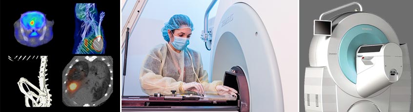
The Siemens Inveon® Multi-Modality System is a versatile platform for pre-clinical CT, SPECT, and PET studies on a single integrated gantry. The system can be configured for PET-CT, SPECT-CT, PET-SPECT-CT or CT only. The large area CT system has a field of view up to 10 cm x 10 cm and resolution down to 20 microns. The PET system can deliver 1.4mm FWHM spatial resolution, and a maximum field of view of 10cm x 30cm. Whole mouse and rat SPECT studies are possible using gamma rays ranging in energy from 30 to 300 keV, with automated zoom for optimising the field of view.
The setting uses a dedicated COBRA reconstruction workstation that runs on a 64-bit multi-core processor to perform real-time reconstruction of an entire mouse within a few minutes. During the reconstructions, the image acquisition workstation is available to perform additional scans, allowing uninterrupted multi-modality imaging and fast throughput. The scanner uses a dedicated sevoflurane inhalation gas anesthesia system, heated imaging chambers and imaging platforms, and the BioVet Physiological Monitoring System to monitor body-temperature, respiration and cardiac activity. A set of two lasers on the outside of the microCT gantry is used to ensure that specimens will be centered within the field-of-view.
In addition to the microSPECT/PET/CT scanner, the radiochemistry suite is fully equipped with a dedicated shielded animal holding room, a well counter, a Packard Cobra II Gamma Counter (model D5010 with 10-detector system (2-inches NaI through-hole crystal detector, 2000 KeV energy range), and a customized animal holding platform for catheter placement and intravenous injection.
PET key features:
- 20 x 20 LSO crystal array detectors
- 10 cm x 12.7 cm transaxial and axial FOV ,
- 1.4 mm FWHM isotropic spatial resolution at the center FOV
- list-mode acquisition, static or dynamic imaging
- 2D and 3D image reconstructions
- wide range of PET radiotracers including 18F-FDG, 18F-NaF, 18F-FLT, 11C-Acetate, in addition to many more investigational molecular imaging probes labeled with 18F, 11C, 64Cu, 62Cu, 68Ga, 89Zr, 13N, 15O and other positron-emitting radionuclides.
SPECT key features:
- two detectors of pixelated NaI(Tl)-crystals
- spatial resolution between 0.8 mm and 1.25 mm with the multi-pinhole mouse-brain collimators
- 3DOSEM or MAP3D iterative reconstructions
- wide range of SPECT radiotracers including 99mTc-Pertechnetate, 99mTc-MDP, 99mTc-MIBI, 67Ga-citrate, 201Tl-chloride, 111In-Octreotide, 111In-Oxine, 123I-NaI, in addition to many more investigational molecular imaging probes.
CT key features:
- 80 W, tungsten anode, 35-80 kVp standard source
- isotropic image resolution of up to 40 µm,
- FOV from 4.4 cm x 4.4 cm up to 10 cm x 10 cm
Nuclear imaging techniques PET and SPECT allow imaging of radiotracer molecules at picomolar concentrations, and they provide uniquely non-invasive, non-toxic, quantitative, longitudinal and functional images of tumor biology. They are also useful in diagnosis and for helping to understand the mechanisms of tumorigenesis. PET is more sensitive than SPECT, whereas the spatial resolution of SPECT is better than that of PET in small-animal imaging.
CT provides a high degree of spatial resolution that is well suited for tumor phenotyping and anatomical detail, and is is generally label-free. However, it lacks molecular specificity.
To complement functional imaging with anatomical detail, it is crucial to coregister molecular data collected through PET or SPECT with the more precise association of signal to anatomical regions shown on CT (or MRI).
For more information, download our Inveon® Multi-Modality System PDF