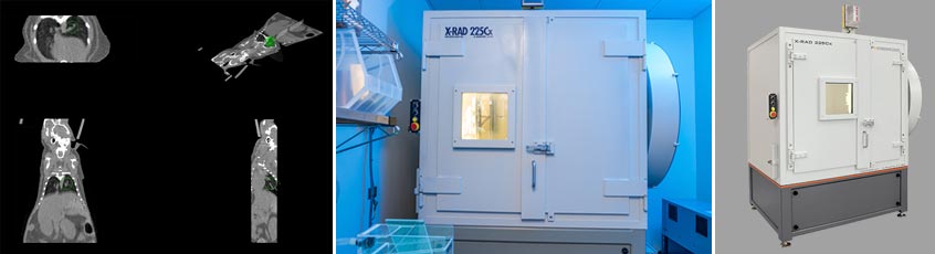
CT image-guided X-ray irradiation is performed on an Precision X-Ray X-RAD 225Cx image-guided biological irradiator System, which is equipped with a 225 kVp X-ray source, pixelated CsI detector. This system is also used to perform cone beam CT imaging, fluoroscopy scans using an Al imaging-beam filter and small focal spot, and image-guided irradiation using the Cu treatment-beam filter and large focal spot. Brass and lead collimator cones are used to define the size and shape the therapeutic beam at the isocenter of the X-ray gantry and the animal platform. The largest “open-field” therapeutic beam can deliver uniform dose to an 11 cm x 11 cm square field at isocenter for whole-body irradiation. The collimator cones can be used to cone-down the therapeutic beam to various sizes ranging from 4 cm through 1 mm, and into circular, square, and rectangular shapes, for whole-brain irradiation and for much smaller targets such as in vivo tumors and in vitro targets such as cell cultures. The X-Rad image-guided targeting can be as precise as ± 0.5 mm for well defined targets, and the system delivers a dose-rate of 4.22 Gy/min, at 5 mm depth in water at isocenter, with the open-field treatment beam.
This X-rad irradiator uses a dedicated isoflurane inhalation gas anesthesia system for in vivo imaging and therapy, and has a lead glass viewing window built into the shielded cabinet for monitoring animals during treatment. A 64-bit multi-core dedicated workstation uses the Image Acquisition and Reconstruction, 3D alignment and Targeting Software for cone beam CT image reconstruction, and for multi-modality image registration and therapy planning.